
The normal lens of the eye is clear (transparent). As a cataract develops, the lens becomes cloudy and this blocks light from entering your eye. Delay in treatment of cataract may lead to blindness and severe pain due to excessive raised pressure in the eye; a condition also known as glaucoma.
Cataract surgery is indicated when the cataract impairs vision to the extent that it interferes with normal daily activities. This involves removing the natural lens of the eye and replacing it with an artificial lens (intraocular lens implant or IOL). The man-made artificial lens is permanent, requires no care, and can significantly improve vision. Cataract surgery is typically an outpatient procedure that takes less than an hour. Most patients are awake during the procedure and need only local anaesthesia. While we treat many straight-forward cataract cases, we are also specialized in treating difficult cataract cases in patients with small pupil, high myopia (short sightedness), post vitrectomy and etc.
The lens is removed in one of the following ways, depending on the type of cataract:
- Phacoemulsification: With this procedure, the doctor uses a device that produces ultrasonic sound waves to break up the cataract into small pieces. The pieces are then suctioned out through a tiny surgical opening. This procedure uses a very small incision.
- Extracapsular extraction: This procedure requires a larger incision because the cataract is not broken into small pieces. The doctor uses an instrument to remove the cataract in one piece through a larger surgical opening. This approach may be used if your cataract has advanced to the point where phacoemulsification can't break up the clouded lens.
 |
 |
||
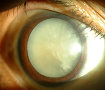
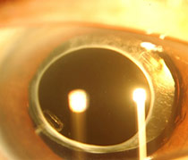
| Pearly white mature cataract caused extremely poor vision | After the removal of cataract, an intra-ocular lens is placed to restore the vision |
With the use of local anaesthesia, the surgery is normally carried out as an out-patient procedure which means it does not require hospitalization. The first step of the procedure is excision of the growth and followed by placing a graft at the area where the pterygium is removed (usually using your own conjunctival tissue). Suturing is often not necessary because the graft could be attached to the eye by using a specialized biocompatible glue. This technique improves surgical wound healing and reduce the chance of growth recurrence.
 |
|
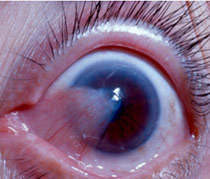
| Fleshy tissue (pterygium) may cover the visual axis and cause permanent damage to the eye sight |